
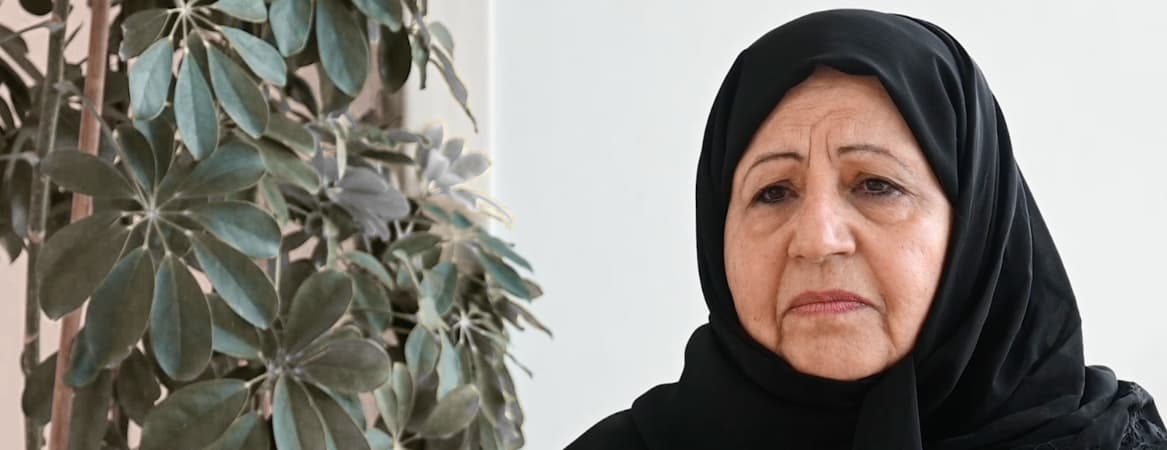
JHAH research suggests AI-enhanced medical imaging can help detect early-stage heart disease in cancer patients
May 12, 2025
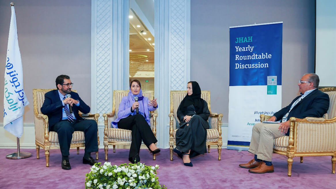
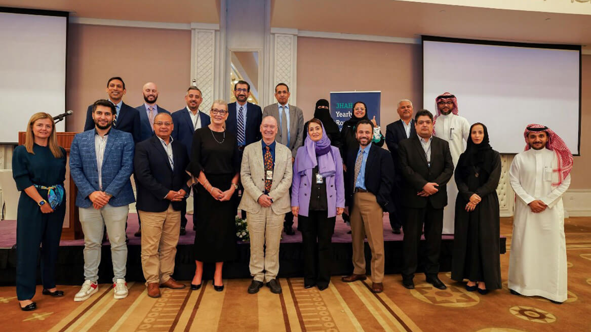
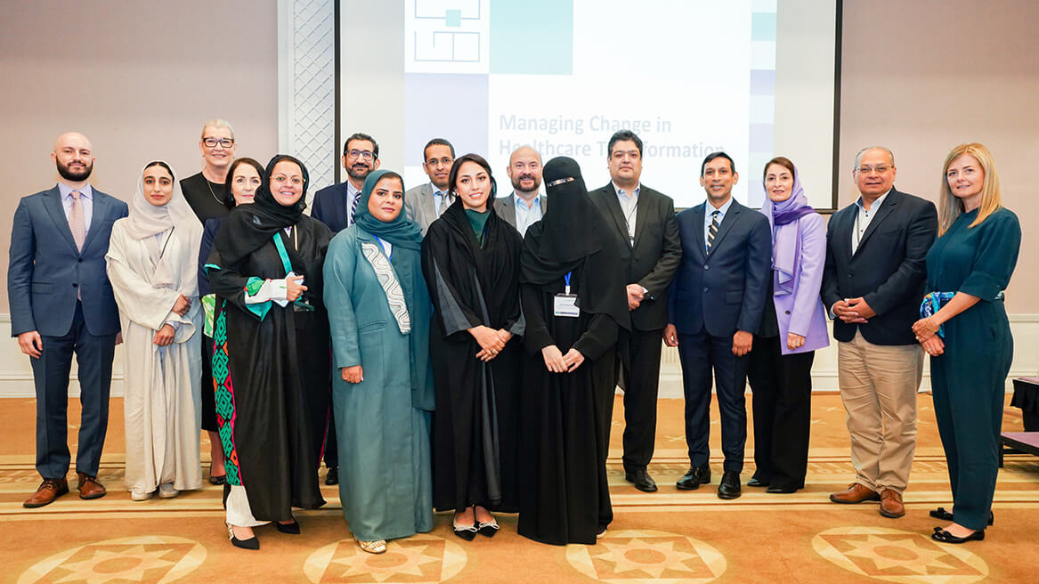
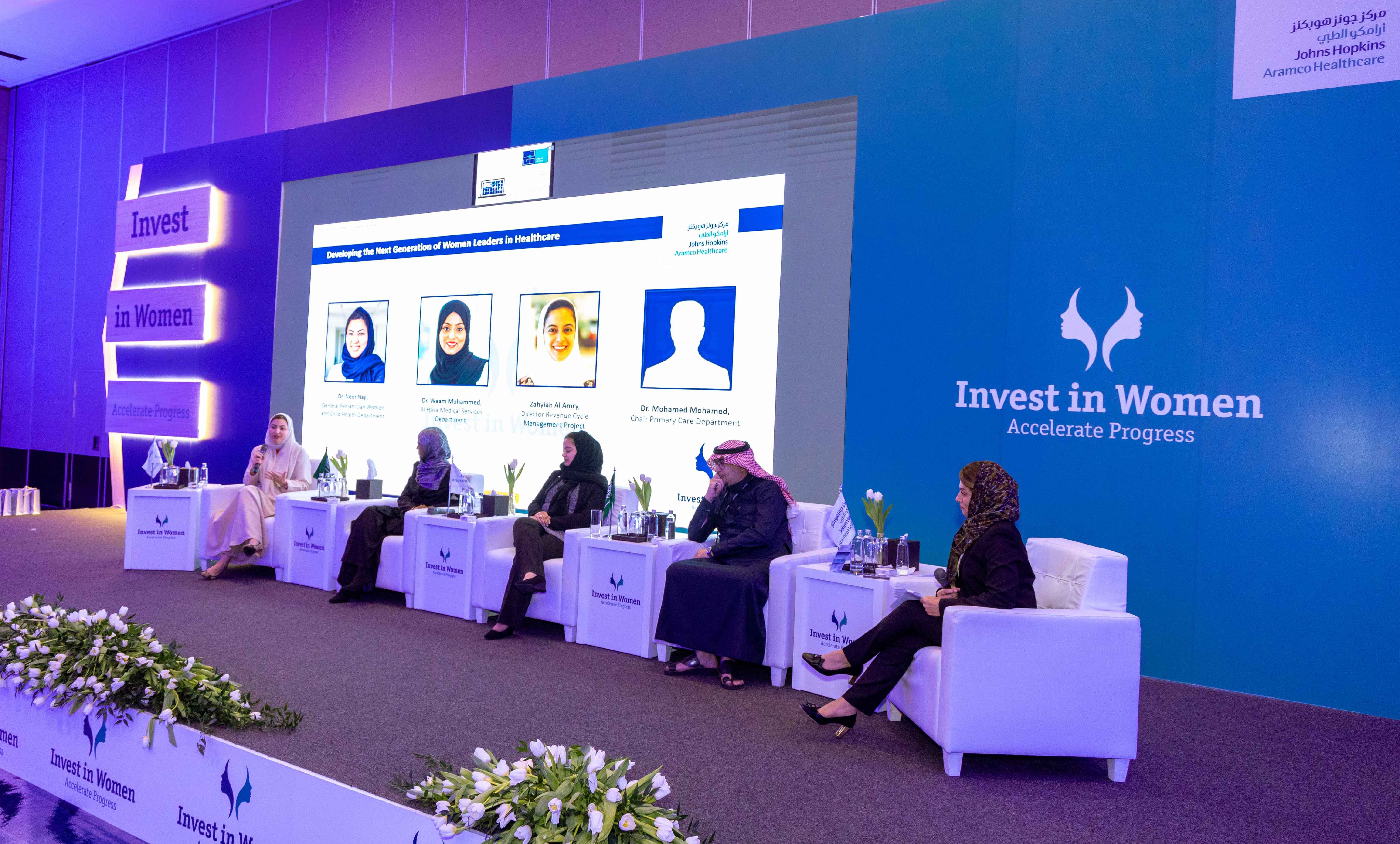
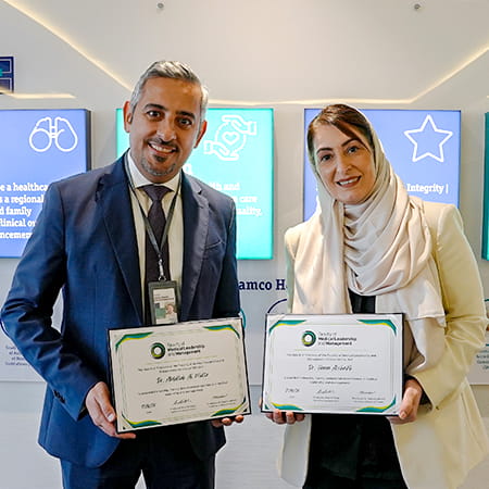
Managing Change in the Healthcare Transformation Process
Johns Hopkins Aramco Healthcare's recent roundtable titled Managing Change in Healthcare Transformation provided an insightful and timely discussion.
This year’s event brought together a diverse panel of experts and thought leaders from around the world to dissect the challenges and opportunities presented by the evolving landscape of healthcare.
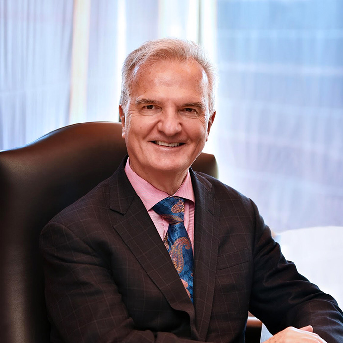
Dr. Michael Walsh, Chief Executive, JHAH, noted the significant transformations in healthcare being driven by technological advancements, shifting demographics and the ongoing global health crisis.
The event comprised renowned healthcare leaders, clinicians and researchers who are playing pivotal roles in shaping the industry as the Kingdom moves to fulfilling its 2030 vision.
One key takeaway was the importance of patient-centered care with an emphasis on patient empowerment and engagement by fostering collaborative relationships between patients and healthcare providers.
The challenges inherent in transforming healthcare were topics of interest. These included healthcare professionals needing to adapt to new workflows and the importance of continuing education and training to ensure healthcare teams are prepared to effectively navigate change.
Collaboration emerged as a central theme throughout the discussion as being vital for successful healthcare transformation.
“Many of the topics were very important, in particular how you can be an effective leader,” said Dr. Ali Zalai, Ministry of Health. “We are learning to deal with the change of mindset.”
“The event showcased a blend of mixed and diverse experiences in managing change in healthcare transformation,” said Mohammed AlAreeky, Advisor, Vision 2030 Health Sector Transformation Program.
“The lessons learned and culture of sharing knowledge is quite pivotal to the success of transformation in the health sector as we shift towards promoting healthier communities and value-based care.”
The roundtable discussion concluded with featured insights on effectively managing change from JHAH physicians Dr. Hanan AlShaikh; Dr. Peter Bibawy; and Dr. Ahmed Jameel; as well as Dr. Dalia Mominkhan, Ministry of Health.
The JHAH Leadership Roundtable provided valuable insight into the evolving healthcare landscape and underscored the necessity for adaptability, technology integration, patient-centered care and collaboration. As JHAH continues to evolve, events such as this are instrumental in facilitating constructive dialogue and driving positive change in healthcare delivery in the Kingdom.
World Breastfeeding Week
July 31, 2023
JHAH joins the World Alliance for Breastfeeding Action and the MOH in supporting World Breastfeeding Week
Leading the way in family medicine
November 19, 2018
We are providing state-of-the-art teaching modalities to help build the skills of future family physicians
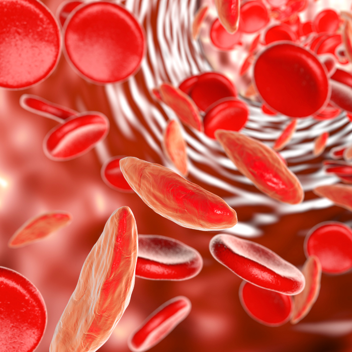
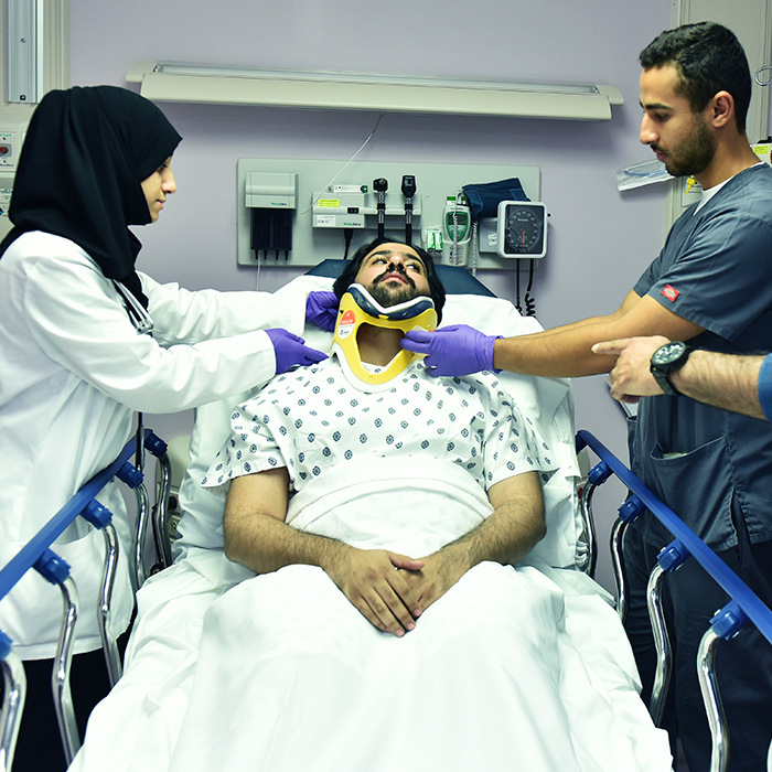
Tackling the pain of sickle cell
November 19, 2018
Find out how changes to how we manage the pain of sickle cell disease have led to a massive drop in emergency care and hospitalization
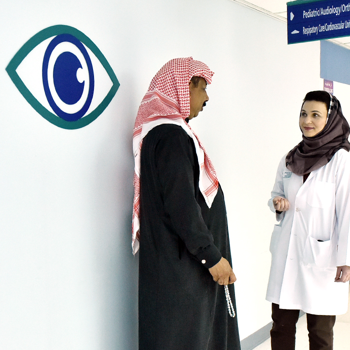
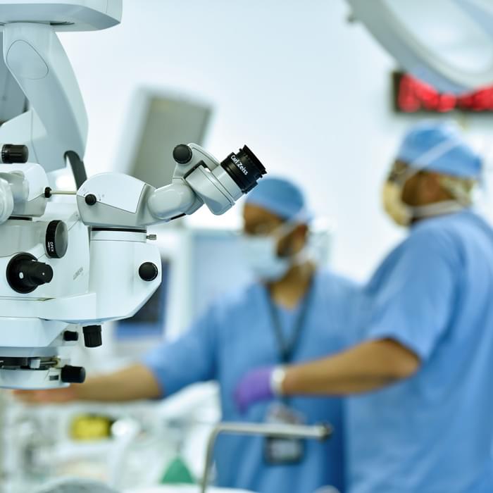
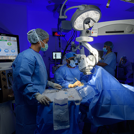
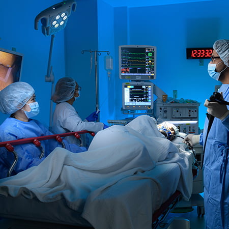
Sight-saving surgery
November 19, 2018
How eye surgery at JHAH has helped save the sight of three local residents
Growing to serve you better - Al-Hasa Health Center
January 17, 2019
Mostly growth is a slow, steady process. Al-Hasa Health Center is currently experiencing a growth spurt

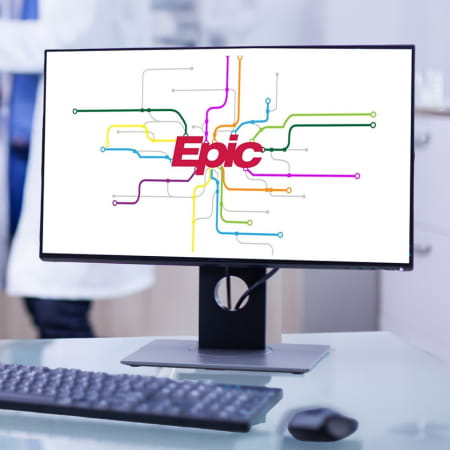
First in Saudi - JHAH launches Epic
November 19, 2018
JHAH is the first hospital in the Kingdom to integrate the leading electronic health record system, Epic
JHAH CEO supports inclusion and diversity
August 01, 2019
JHAH CEO outlines his commitment to providing an inclusive, adaptive environment for everyone, including patients, families, visitors, and employees
Accredited for Postgraduate Medical Training
October 26, 2019
JHAH Becomes the First Private Institution to be Accredited by Saudi Commission for Health Specialties (SCFHS) for Postgraduate Medical Training
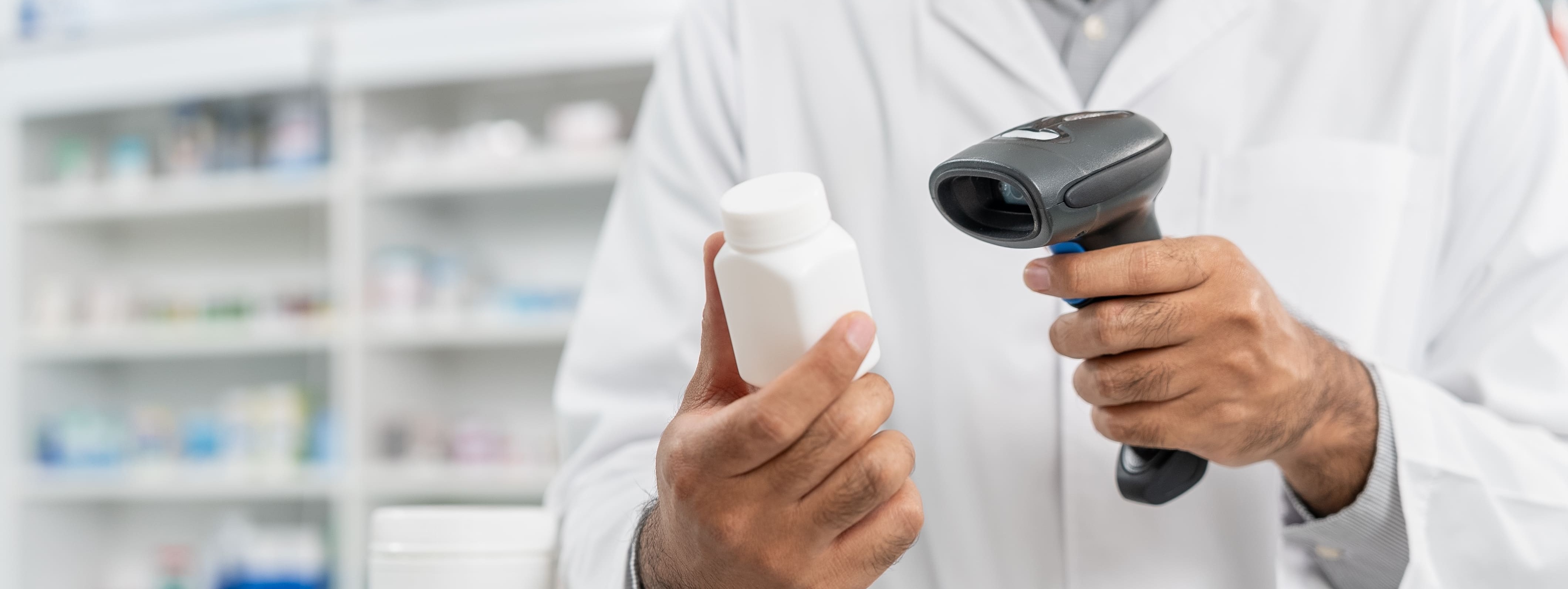
JHAH Pharmacy Wins Patient Safety Award
November 19, 2019
JHAH presented with the 2019 National Patient Safety Award for a project in the category “Medical Institutions / Pharmacy”.
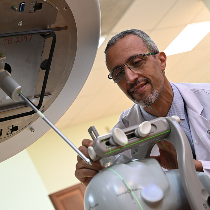
Stereotactic Radiosurgery
January 29, 2020
JHAH is proud to be the first hospital in the Eastern Province to conduct a Stereotactic Radiosurgery using TrueBeam accelerators
Person-Centered Care Certification
April 08, 2020
JHAH awarded “Gold” Person-Centered-Care Certification by Planetree
Echocardiography Accreditation
June 21, 2021
One of only two organizations in the Kingdom, and one of three in the GCC, with accreditation in Echocardiography - Adult Transthoracic
JHM experts’ life-saving impact
September 22, 2021
Specialists from Johns Hopkins Medicine U.S. are sharing their expertise in ongoing medical rotations on the ground at JHAH

JHAH Actively Supports Inclusion and Diversity
October 01, 2023
JHAH Chief Operating Officer (COO) outlines his commitment to providing an inclusive, adaptive environment for everyone

How a JHAH Physician Saved a Life
May 10, 2022
A Saudi Aramco employee thanks his physician at JHAH for successfully removing his brain tumor
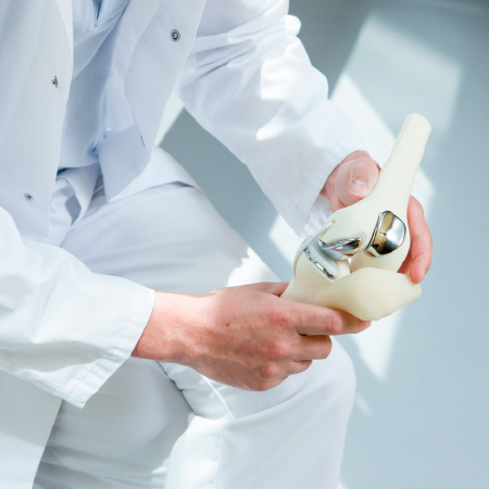
A Total Hip Replacement Relieves Patient's Pain
June 19, 2022
JHAH Orthopedic team performed a complex surgery that significantly reduced the chronic hip pain of a patient born with a rare case
Total-Knee Replacement in the Kingdom
October 04, 2022
The misconceptions and misinformation over a globally adopted and life-changing surgery are changing; learn more
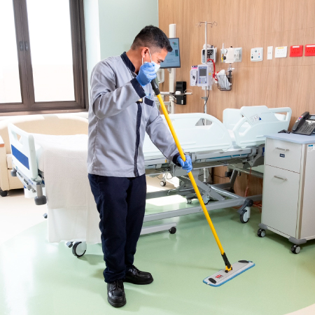
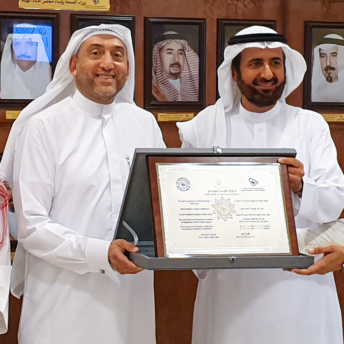
JHAH Achieves CIMS Accreditation
November 16, 2022
JHAH achieved the ISSA’s Cleaning Industry Management Standard (CIMS) certification with honors.
Improving Patient Outcomes Using Artificial Intelligence (AI)
December 07, 2022
How JHAH is Using AI Cognitive Computing Models to Identify Deteriorating Patients Sooner
Imaging Skills at JHAH
December 15, 2022
The (MRI) team utilized their imaging skills to confirm the success of a lifesaving. Read more
Arthroscopic Subtalar Surgery
December 25, 2022
JHAH is a regional pioneer in using MIS on the foot and ankle. Read more
Esophageal pH and Manometry
December 27, 2022
Dr. Diamond Joy, Gastroenterologist, and team performed the first pH of manometry service and the first case of adult esophageal manometry at JHAH

JHAH Leadership Roundtable 2023
September 10, 2023
Johns Hopkins Aramco Healthcare's recent roundtable titled Managing Change in Healthcare Transformation provided an insightful and timely discussion
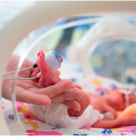
World Prematurity Day
November 17, 2022
November 17, is a global awareness day that aims to draw attention about preterm birth

A common problem for men, easily solved
January 07, 2024
JHAH introduced Rezum Water Vapor Therapy to quickly and easily solve the problem of enlarged prostates
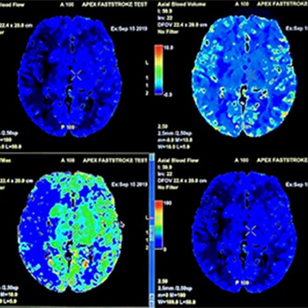
Advanced Radiology Diagnostic Procedure at Al-Hasa
December 29, 2023
Al-Hasa Health Center (AHHC) is now offering CT perfusion, a specialized-ray that uses computed tomography to show the flow of blood in the brain.

Al-Hasa Pharmacy Participates in Antimicrobial Week
January 01, 2024
JHAH Al-Hasa Pharmacy participated in a Global Antimicrobial Week event held at King Faisal University in coordination with the Ministry of Health’s Public Health Authority

Art from the Heart
January 01, 2024
For more than eleven years, the Dhahran-Aramco Girl Scout Troop 22 has supported cancer patients, particularly children, and their families. This year they saw an opportunity and took it as their own
Ada'a Health Program Award
February 26, 2024
JHAH has won the Ada'a Health Program Award for a clinical audits project on Improving Compliance with Venous Thromboembolism (VTE) Risk Assessment and Prophylaxis
JHAH Orthopedics Performs a Surgical First
March 06, 2024
The JHAH Orthopedics Surgery Department added another treatment option to the already impressive list of options it provides patients suffering from serious ankle pain.

A Streak of Gold
March 19, 2024
Johns Hopkins Aramco Healthcare has maintained gold Mowaamah certification since 2019 with 99% compliance rate

Care Anywhere Continues to Expand
March 20, 2024
JHAH’s Care Anywhere 24/7 access to Primary Care now includes pediatric primary care experts with immediate access
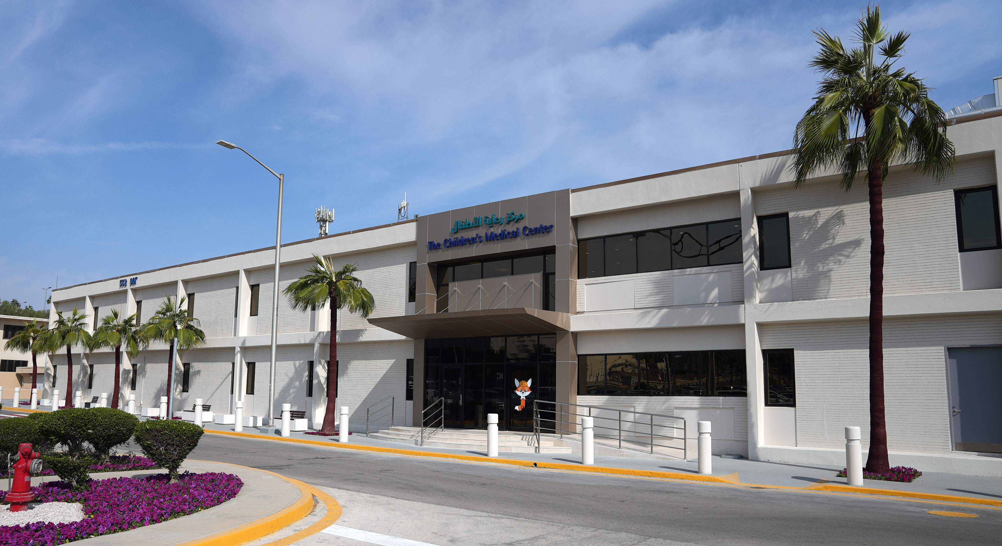
Dhahran Pediatric Specialty Clinic Moved to Building 552
March 20, 2024
The new Children’s Medical Center in Dhahran continues to expand with Pediatric Specialty Services moving to the second floor, Building 552
JHAH Celebrates International Women's Day
March 21, 2024
JHAH joined Saudi Aramco in hosting over 170 healthcare professionals at a special event for International Women’s Day
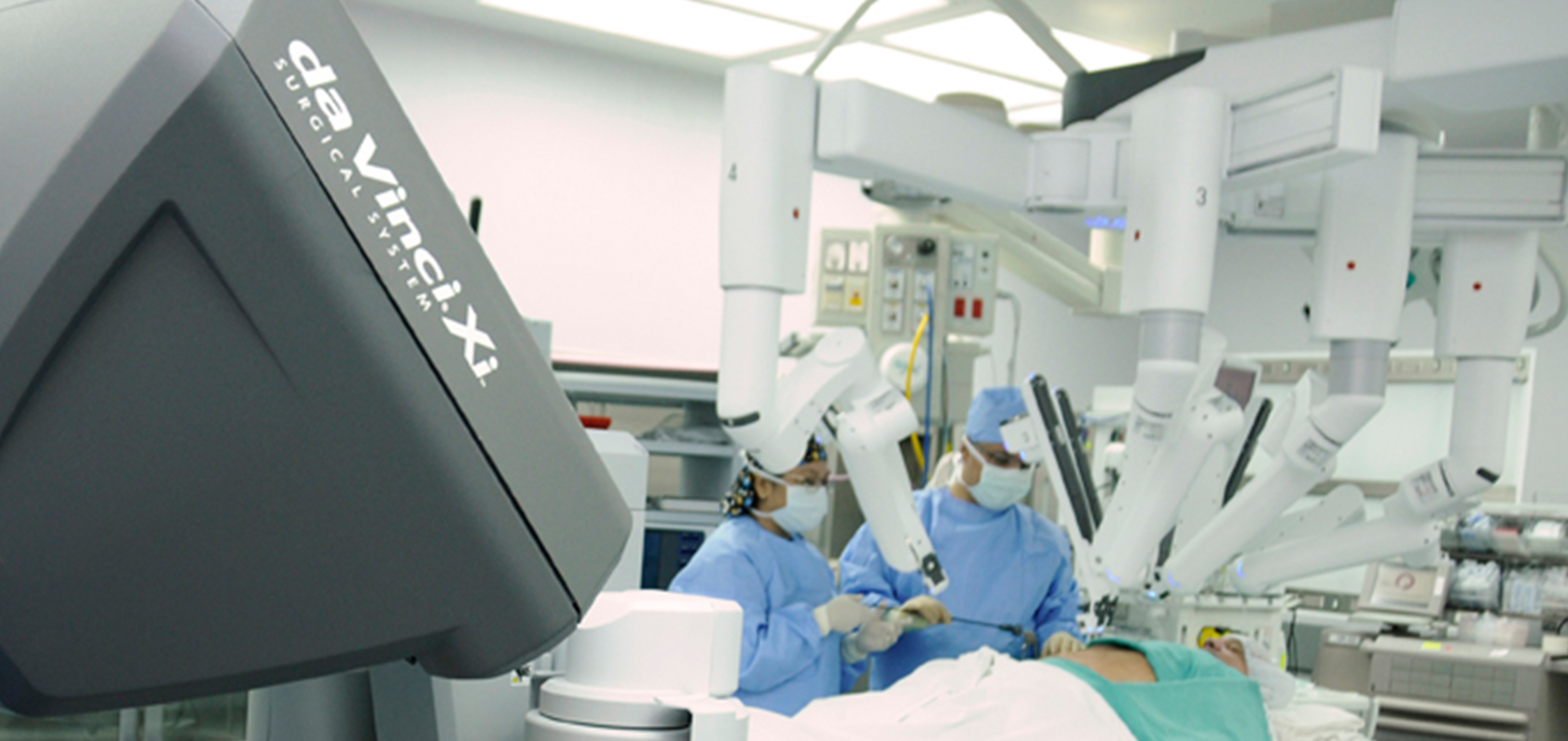
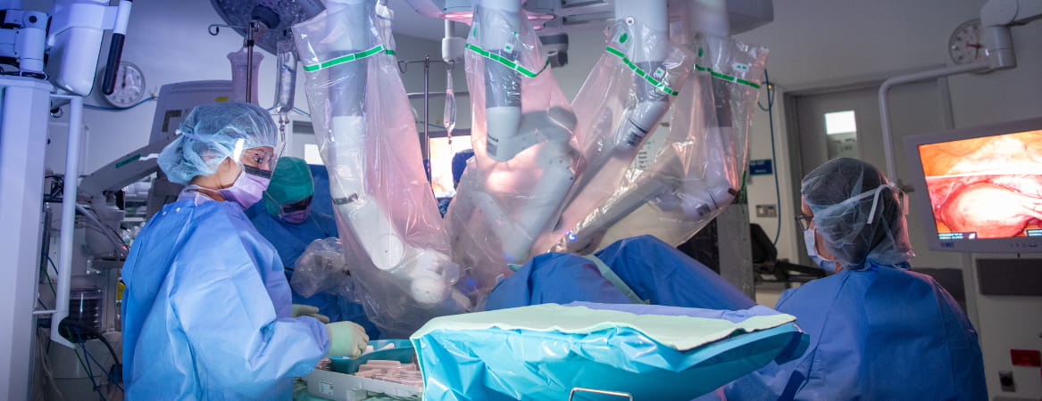
JHAH Robotic-Assisted Surgeries Reach Major Milestone
April 03, 2024
DaVinci Robotic Assisted Surgery achieved a record 300 procedures in 2023
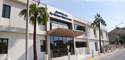
A Behind-the-scenes look at 552
April 03, 2024
Dhahran Children’s Medical Center highlights our commitment to quality and safety of care
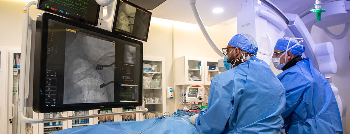
First with Pulsed Field Ablation Technology
May 05, 2024
Johns Hopkins Aramco Healthcare becomes first facility in Eastern Province to implement “Pulsed Field Ablation Technology”

Another JHAH Regional First
May 14, 2024
JHAH is the first healthcare institution in GCC to obtain Pediatric and Congenital Transthoracic Echo Lab Accreditation

Specialized Eye Care Now Available to Ras Tanura Patients
May 15, 2024
No more monthly trips to Dhahran Medical Center to receive Vascular Edothelial Growth Factor injection
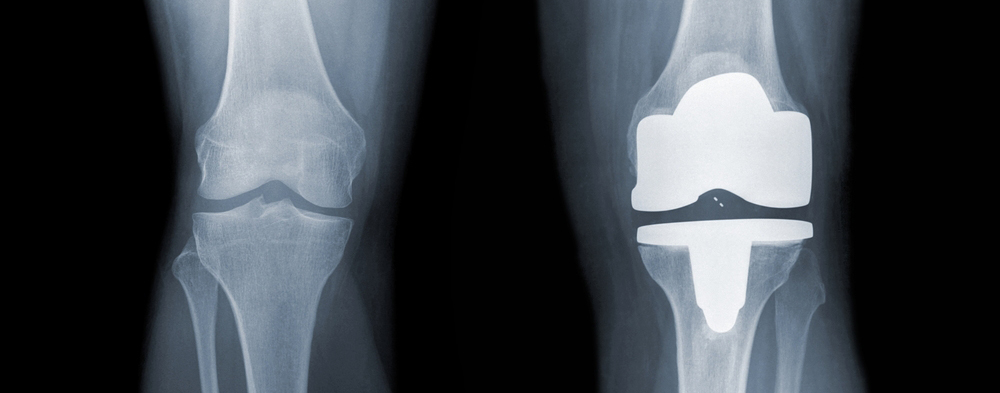
Total Knee Replacement: A Lifechanging Procedure
May 15, 2024
Al-Hasa Health Center Orthopedics dramatically increases Total-Knee Replacement surgeries

JHAH Shines at MEFMA 2024
May 14, 2024
JHAH Facilities Management & Services Team wins Client Best Practices (Corporate) and Digital Transformation Awards.

JHAH Inaugural Chairman Award Ceremony
May 15, 2024
Celebrating the excellence of JHAH individuals and teams for their positive effect on patient care and the organization
A Healthy Spine: The New JHAH Scoliosis Clinic
May 15, 2024
Discover dedicated scoliosis care—from diagnosis to treatment—at the new JHAH Scoliosis Clinic

A Gift of Love to JHAH Newborns
July 11, 2024
Dhahran Oasis Quilt Guild has taken dedication to an entirely new level in guild’s 21st anniversary.

A Parent’s Nightmare: A JHAH Emergency Room Response
July 15, 2024
Our registered nurse Fraulein Tabuzo saves baby Arwa
A New Inventory Management Solution Goes Live
July 15, 2024
The Supply Chain Department optimizes inventory management with an innovative state-of-the-art barcoding system
Another JHAH First: Tongue and Mouth Reconstruction
July 15, 2024
JHAH team of surgeons conduct the first complex resection and reconstruction of the tongue and mouth floor
Another JHAH First: Robotic Kidney Stone Removal
July 16, 2024
JHAH urological surgery team conducted its first robotic pyelolithotomy of a kidney stone in an abnormally positioned kidney
Autologous Hematopoietic Stem Cell Transplantation Program (AHSCT)
September 11, 2024
A vital treatment option for multiple myeloma, lymphoma and other cancers

Join Us in Protecting the Earth
September 15, 2024
Recycling Guidelines at JHAH to safeguarding the environment
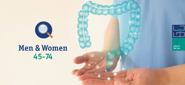
Colon Cancer Screening: A Crucial Step for Your Health
September 16, 2024
Colon cancer screening can detect the disease in its early and treatable stages
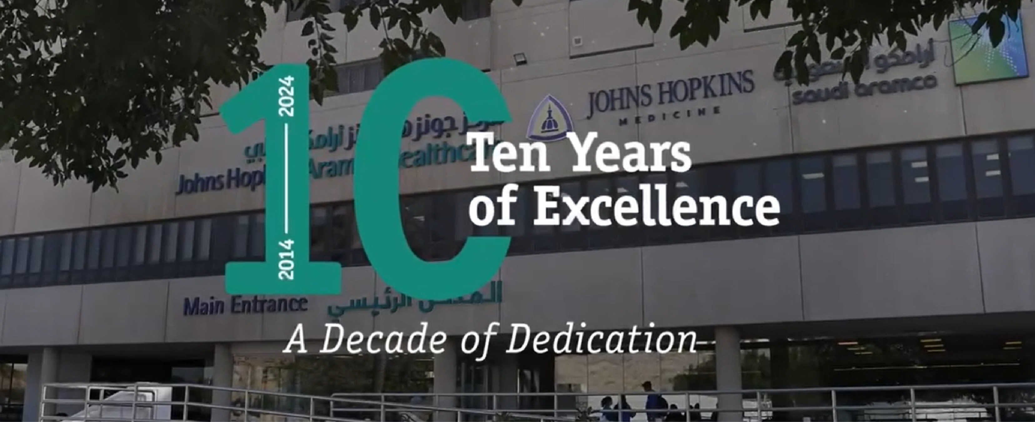
A Decade of Dedication
November 05, 2024
Marking a decade, Saudi Aramco and Johns Hopkins Medicine renew their commitment to world-class healthcare.
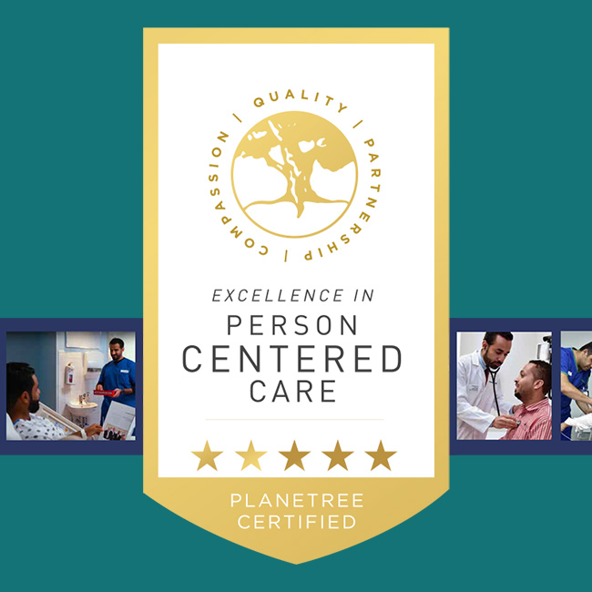
JHAH Receives Highest Certification for Excellence in Person-Centered Care from Planetree
November 12, 2024
Johns Hopkins Aramco Healthcare (JHAH) Receives Highest Certification for Excellence in Person-Centered Care from Planetree

Person-Centered Care Earns JHAH Prestigious Award Again
November 21, 2024
Person-centered care helps JHAH attain second consecutive prestigious award
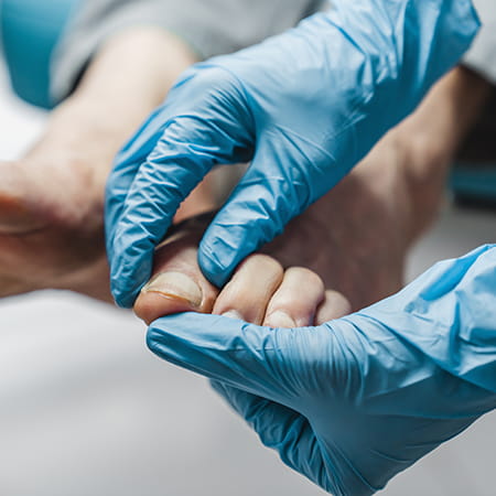
Podiatry's Vital Role in Diabetes Care at JHAH
November 26, 2024
Explore how JHAH’s podiatry experts transform diabetic foot care and protect mobility this World Diabetes Month!
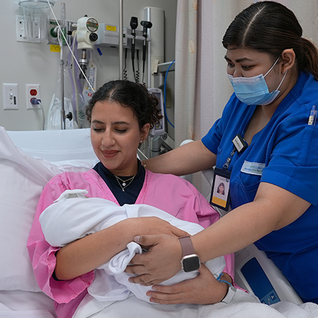
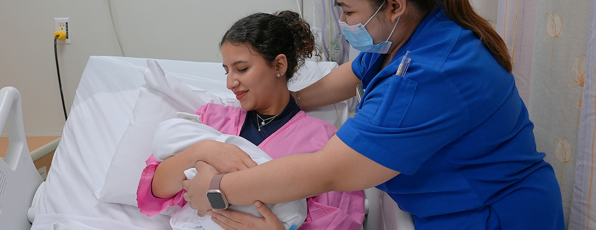
JHAH Sets New Standard in Maternal Care
November 28, 2024
Leading the way in maternal care, JHAH empowers families with expert support and a commitment to breastfeeding excellence
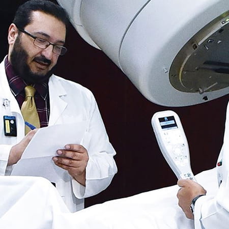
Meet the two JHAH doctors recognized as “pioneers” of radiotherapy in Saudi Arabia
December 05, 2024
Explore the stories of two JHAH doctors shaping the future of radiotherapy in Saudi Arabia with groundbreaking technology and heartfelt dedication
JHAH performs first keyhole surgical procedure of its kind in Eastern Province to correct foot deformity
December 15, 2024
Discover JHAH's groundbreaking keyhole bunion correction surgery in the Eastern Province, offering faster recovery, less pain, and immediate mobility after the procedure
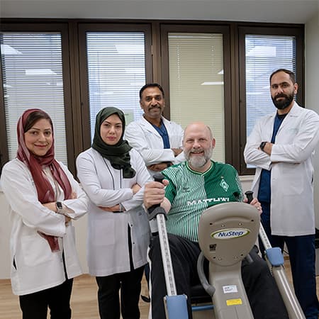
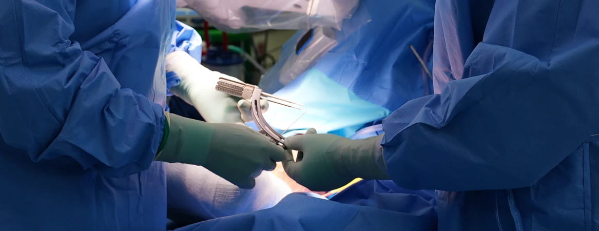
From heart attack to better health: A patient’s journey through JHAH’s first-of-its-kind cardiac rehab program
December 19, 2024
From a heart attack to better health, Greg Noakes shares his recovery journey through JHAH’s first-of-its-kind cardiac rehab program

Meet the 9-year-old JHAH patient whose diabetes diagnosis inspired him to launch a YouTube channel
December 19, 2024
MQ’s diabetes journey, from diagnosis to advocacy, showcases the power of care, family, and determination at JHAH
How JHAH neurosurgeons saved a 16-month-old boy’s life by removing his brain cyst
December 19, 2024
Learn how JHAH neurosurgeons’ swift intervention saved 16-month-old Husain Ali Al Mahdi’s life, giving him a second chance at growth and health
JHAH leaders awarded prestigious fellowships for groundbreaking domestic violence, COVID-19, and patient access initiatives
December 30, 2024
Dr. Hanan Al Shaikh and Dr. Abdullah Al Mulla receive UK fellowships for leading groundbreaking healthcare initiatives at JHAH.

Perfect pass rate for JHAH's family medicine residents
December 31, 2024
JHAH’s Family Medicine Residency Program celebrates a perfect 100% pass rate for its 2024 final-year residents, setting a new standard for primary care

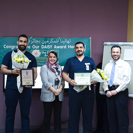
Celebrating Excellence: JHAH Nurses Honored with DAISY Awards for Extraordinary Care
January 08, 2025
Join us in celebrating JHAH’s DAISY Award-winning nurses, Hebzibaa, Mohrah, and Zubida. Read their inspiring stories and nominate your favorite nurse!
A patient’s 16-year journey with JHAH’s retina surgeons brings clarity of vision for their retirement
January 14, 2025
From struggling with thick contact lenses to crystal-clear vision: one patient's transformative journey with JHAH.
JHAH Al-Hasa unveils two new services for gastroenterology and dialysis patients
January 19, 2025
First private hospital in Al-Hasa to offer automated peritoneal dialysis and one of only a few offering ERCP.
JHAH Dhahran’s Emergency Department reports record patient satisfaction in 2024 as visits hit all-time high
January 19, 2025
Patient visits double while satisfaction reaches record 81% at JHAH Dhahran's Emergency Department
JHAH Nursing Teams Win DAISY Award: Transforming Cancer Care Through Compassion
January 28, 2025
A young mother facing cancer found strength through her dedicated nurse coordinator. This is just one story behind JHAH's DAISY Award-winning teams.
From Patient to Nurse: A Breast Cancer Survivor’s Inspiring Journey
February 04, 2025
Gitu Mirchandani’s journey from patient to aspiring oncology nurse shows how hope can inspire healing in others
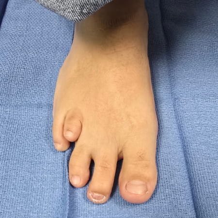
JHAH becomes first hospital in Eastern Province to offer routine toe-lengthening surgery for brachymetatarsia
February 18, 2025
JHAH is the first hospital in the Eastern Province to offer routine toe-lengthening surgery for brachymetatarsia. Discover how this corrective procedure can relieve pain, improve mobility, and restore confidence.
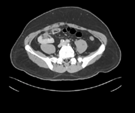
JHAH surgeon conducts first known robotic surgery of its kind in Saudi Arabia to treat colon cancer patient
February 23, 2025
JHAH successfully performed Saudi Arabia’s first robotic colon cancer surgery using an advanced minimally invasive technique, improving patient recovery and outcomes.
Home Healthcare Program at JHAH
December 08, 2022
The program covers a 50 kilometer radius of JHAH’s healthcare facilities. Read more about it..
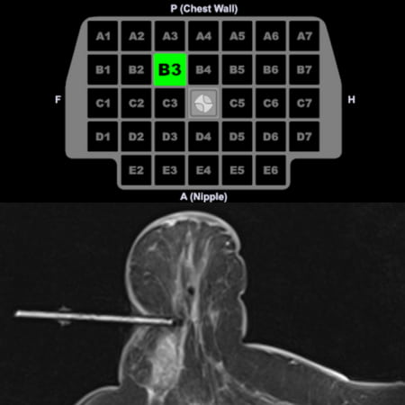
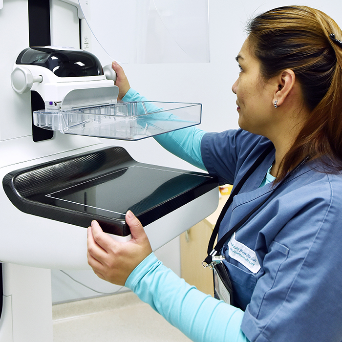
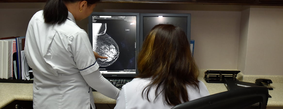
JHAH unveils precision imaging technology for breast cancer screening and diagnosis
March 13, 2025
JHAH expands breast cancer screening with MRI-guided biopsy and contrast-enhanced mammography, improving diagnostic accuracy and patient care
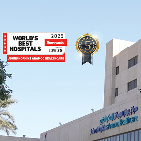
Johns Hopkins Aramco Healthcare Ranked 5th in Saudi Arabia’s Newsweek Best Hospitals 2025 Report
March 20, 2025
Discover how Johns Hopkins Aramco Healthcare rose rapidly to become Saudi Arabia's 5th-best hospital in Newsweek's prestigious 2025 ranking, delivering exceptional patient care and clinical innovation.
Thanks to Hospital at Home, we are able to live in peace
March 20, 2025
Discover how JHAH’s Hospital at Home brings hospital-quality care directly to patient's homes, enhancing comfort and wellbeing.
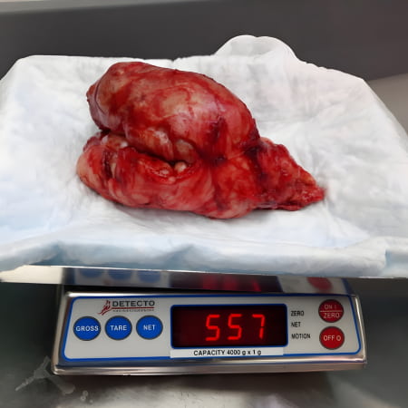
JHAH surgeon removes “huge” prostate weighing more than half a kilogram
April 29, 2025
Discover how JHAH’s expert urology team tackled a rare, extreme case of prostate enlargement with lifesaving surgery and patient-centered care
JHAH first in Middle East to use Epic Dorothy to enhance home healthcare with digital automation
May 01, 2025
JHAH is transforming home healthcare with a digital tool that gives clinicians more time with patients and less time on paperwork
Quick and easy eye test for young children now available at JHAH Dhahran
May 01, 2025
Discover how JHAH’s pediatric team is making early screening for vision and bilirubin issues easier, faster, and more accessible for children.

Children’s Medical Center enhances care for kids with autism and their families by becoming a Certified Autism Center™
May 08, 2025
JHAH’s Children’s Medical Center earns Certified Autism Center™ designation, ensuring a sensory-friendly, supportive environment for children with autism and their caregivers.
Nurses & Midwives Week 2025: A Message from JHAH’s Chief Nursing Officer
May 11, 2025
Read the heartfelt message from JHAH’s Chief Nursing Officer and see why Nurses & Midwives Week 2025 is all about honoring your extraordinary impact.
JHAH becomes first hospital in Eastern Province to receive world-class accreditation with distinction for nursing residency program
May 15, 2025
JHAH is now the first hospital in the Eastern Province to receive international accreditation with distinction for its nurse residency program.
Cutting-edge endoscopic and diagnostic technology help JHAH’s gastroenterology unit to dramatically reduce patient wait times
May 18, 2025
New technology has transformed gastroenterology care at JHAH, cutting routine wait times and improving patient outcomes with less invasive, faster procedures.

JHAH surgeon discovers glass shard in patient’s eye that went undetected for nearly 15 years
May 18, 2025
An extraordinary discovery by a JHAH eye surgeon brings 20/20 vision and lasting relief to a patient who unknowingly lived with a glass shard in his eye for 15 years.
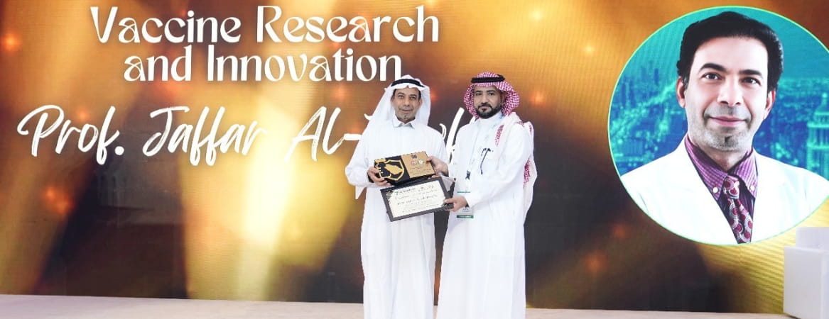
JHAH’s Prof. Jaffar Al-Tawfiq Wins Saudi Vaccine Award for Research on Coronavirus and Hajj Safety
May 28, 2025
Prof. Jaffar Al-Tawfiq was honored for his impactful research into vaccine development, pandemic response, and protecting public health during large-scale events like Hajj.
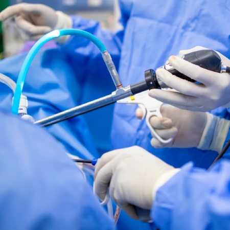
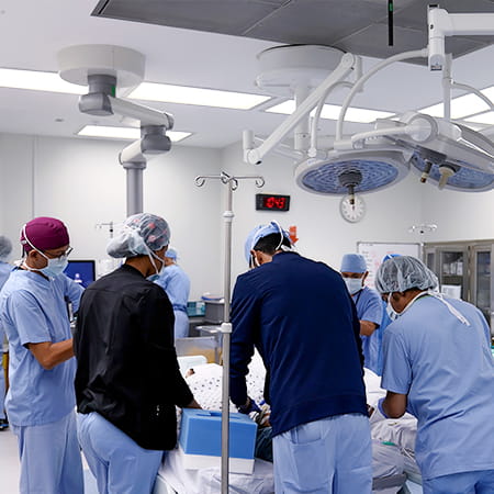
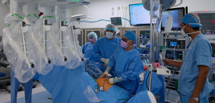
JHAH conducts first robotic rectal surgery of its kind in Eastern Province
June 22, 2025
A JHAH surgical team successfully used robotic Transanal Minimally Invasive Surgery (TAMIS) to remove a rectal tumor with no incisions, marking a new milestone in minimally invasive colorectal surgery.

Three JHAH family medicine physicians scoop national awards
June 19, 2025
JHAH celebrates three family medicine physicians recognized at the national level for clinical excellence, leadership, and impact on healthcare education in Saudi Arabia.
‘Diabetes doesn’t stop me doing anything’: Meet the 15-year-old who is confronting diabetes head-on
July 01, 2025
Diagnosed with diabetes during Ramadan, Rashid turned a health crisis into a story of strength, healing, and hope—thanks to expert care at JHAH.
New Laser Technology Helps JHAH Urologists Zap Kidney Stones with Greater Precision
July 08, 2025
Discover how the advanced MOSES™ laser is transforming kidney stone treatment at JHAH—faster procedures, fewer complications, and a quicker recovery for patients.

‘Like a Hotel’: Room Service Now Available in all JHAH Inpatient Wards
July 08, 2025
Inpatients at JHAH can now enjoy hotel-style room service—customized meals, flexible timing, and easy ordering through MyChart or a quick phone call.
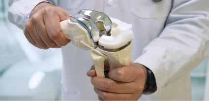
3D Knee Replacement at JHAH Restores Patient's Mobility in Just 24 Hours
July 21, 2025
A breakthrough surgery at JHAH helped a patient walk unaided and climb stairs the very next day. Explore how 3D technology is reshaping knee replacement recovery.

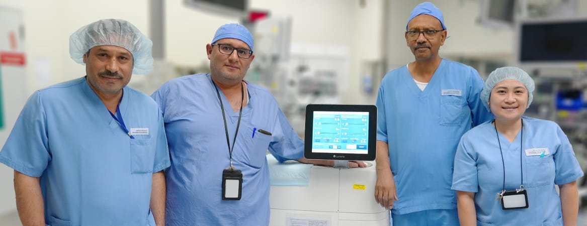
JHAH Cardiologists and Surgeons Beat US Benchmarks for Minimally Invasive Aortic Valve Replacement
July 22, 2025
Exceeding U.S. outcomes, right here in Dhahran. See how JHAH is reshaping what’s possible in heart valve care with minimally invasive advanced transcatheter aortic valve replacement (TAVR).

JHAH Completes First Minimally Invasive Nipple-Sparing Mastectomy in Eastern Province
July 23, 2025
At JHAH, women with breast cancer now have access to a groundbreaking, less invasive surgery that supports faster healing, preserves appearance, and empowers recovery without compromise

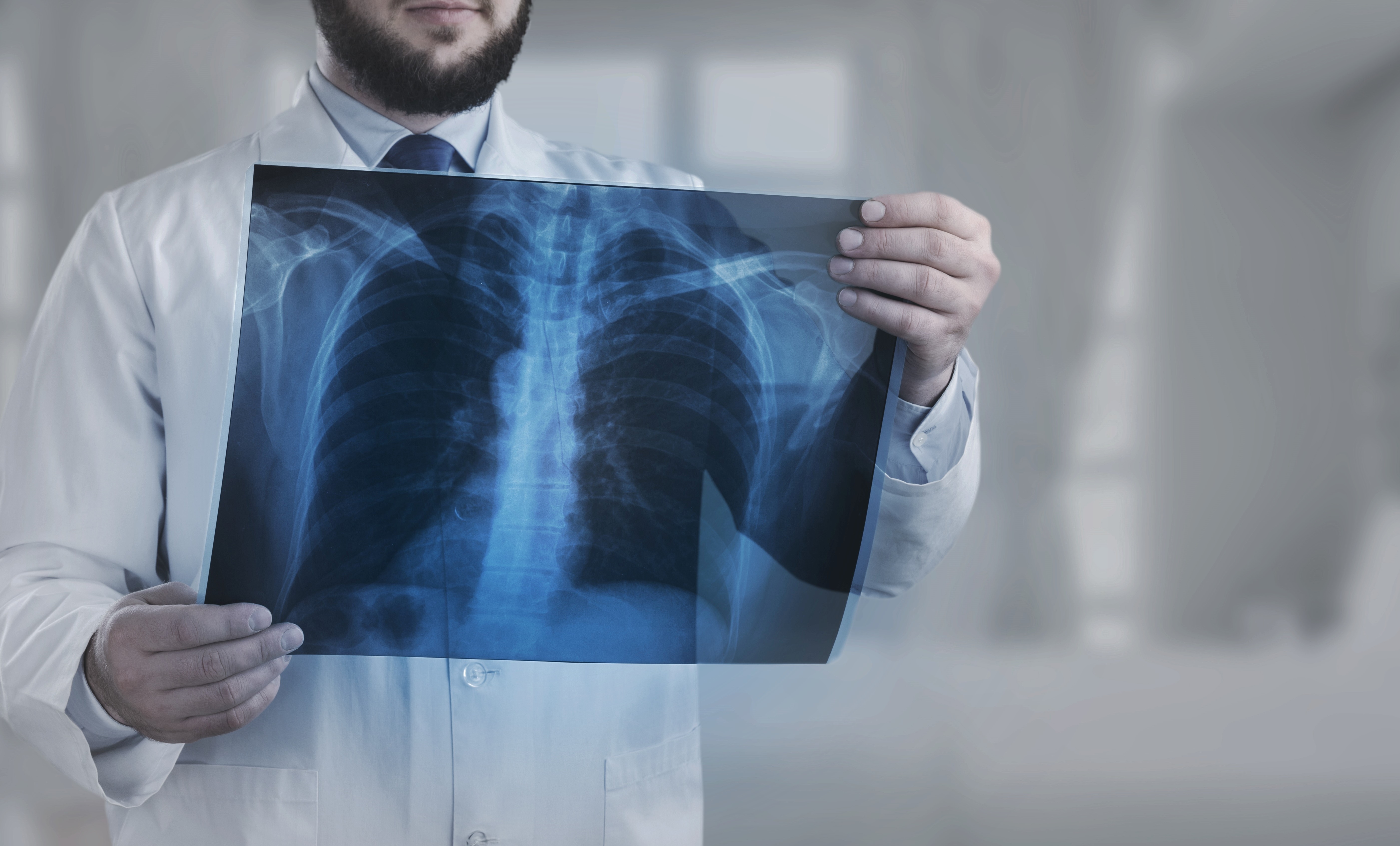
Lung Cancer Patient at JHAH Urges Smokers to Quit Before It’s Too Late
July 31, 2025
After smoking three packs a day for decades, Aisha now urges others to quit. Read how her cancer journey at JHAH is helping raise awareness on World Lung Cancer Day.
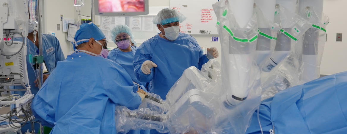
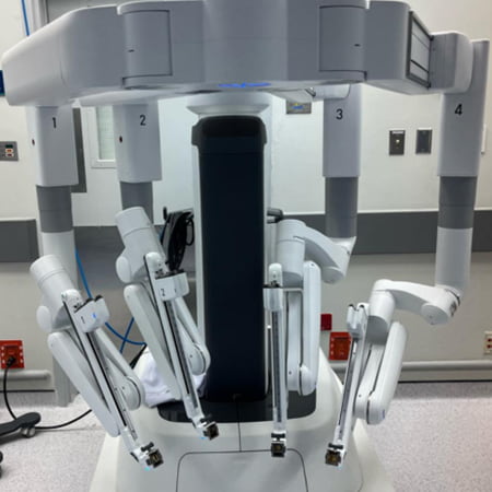
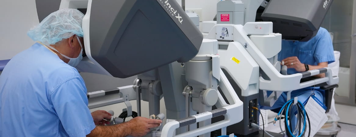
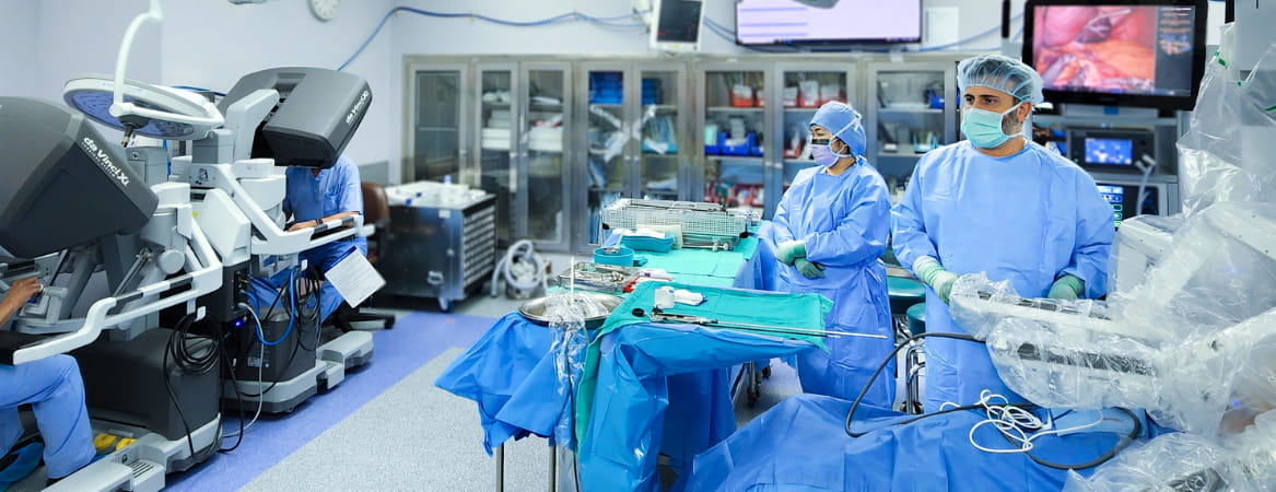
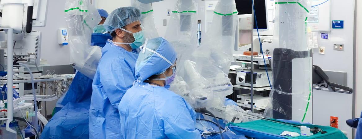
Meet JHAH’s da Vinci surgical robot, the busiest in Saudi Arabia
August 04, 2025
From life-changing heart procedures to intricate cancer surgeries, JHAH’s da Vinci robot is transforming patient care with precision and faster recovery.

‘Sweet, funny, and seriously tough’: The six-year-old girl beating acute leukemia against the odds
September 01, 2025
A six-year-old girl overcame a childhood leukemia relapse with expert care at JHAH. Discover her inspiring survival story of hope and resilience.
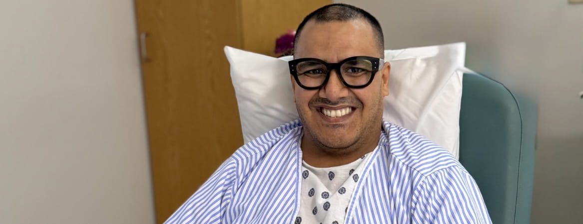
‘Please help me with this pain’: A patient’s journey through a double hip replacement at JHAH
August 24, 2025
At just 41 years old, Wael faced crippling hip pain from a rare congenital condition. After two complex hip replacements at JHAH, he is pain-free and has regained his freedom of movement.
A Patient’s Journey Through Minimally Invasive Heart Surgery at JHAH
August 26, 2025
Fawzia faced severe aortic stenosis but avoided open-heart surgery thanks to JHAH’s minimally invasive TAVR procedure. Discover how she recovered quickly and regained her quality of life.

Helping Clinicians, Advancing Cancer Care: JHAH Radiation Physicist’s Global Training Mission
August 27, 2025
See how JHAH’s Bilal Jalal is empowering clinicians in developing countries through IMRT/VMAT training, transforming cancer care for patients around the globe.
JHAH’s Adult Heart Surgery Program Awarded Elite Status, Ranked in Top 5% Globally
September 22, 2025
Elite recognition, safer outcomes, JHAH’s adult heart surgery program ranks among the world’s best with an STS 3-star rating, the highest possible
JHAH Conducts First Robotic Hernia Surgery of its Kind in Saudi Arabia
September 22, 2025
Saudi Arabia’s first robotic ventral hernia repair with diastasis recti reconstruction is here. See how JHAH surgeons are transforming recovery times and patient outcomes
JHAH Introduces Innovative Erectile Dysfunction Treatment
September 22, 2025
Breakthrough men’s health treatment at JHAH, PRP therapy for erectile dysfunction restores blood flow, improves function, and can be completed in just 20 minutes
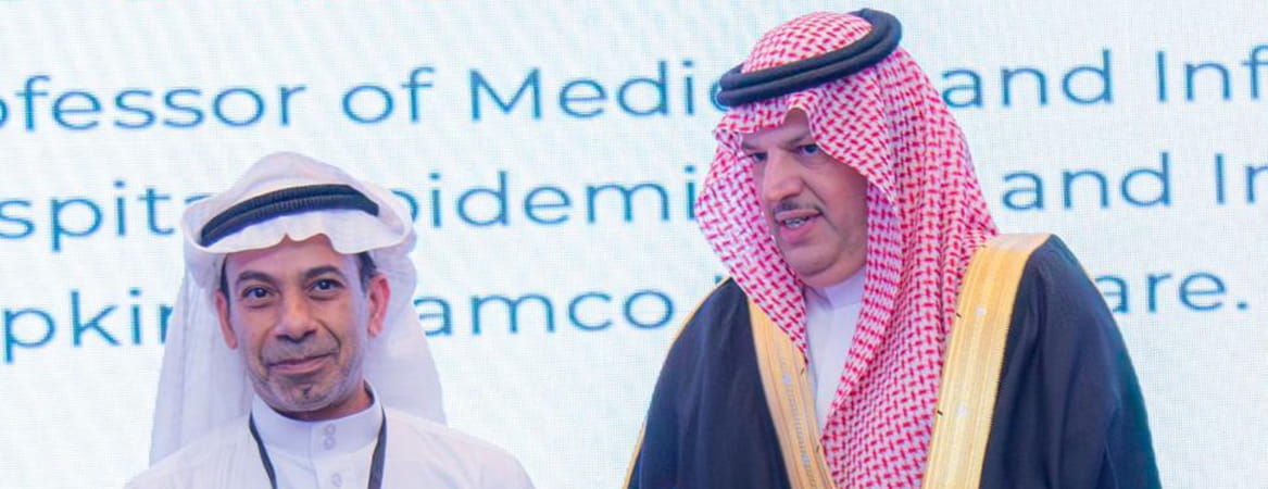
JHAH Infectious Diseases Expert Honored for Global Impact of Clinical Research
September 28, 2025
Global spotlight on JHAH: Prof. Jaffar Al-Tawfiq receives the SPIDS Award for pioneering research that shaped global infectious disease preparedness and response
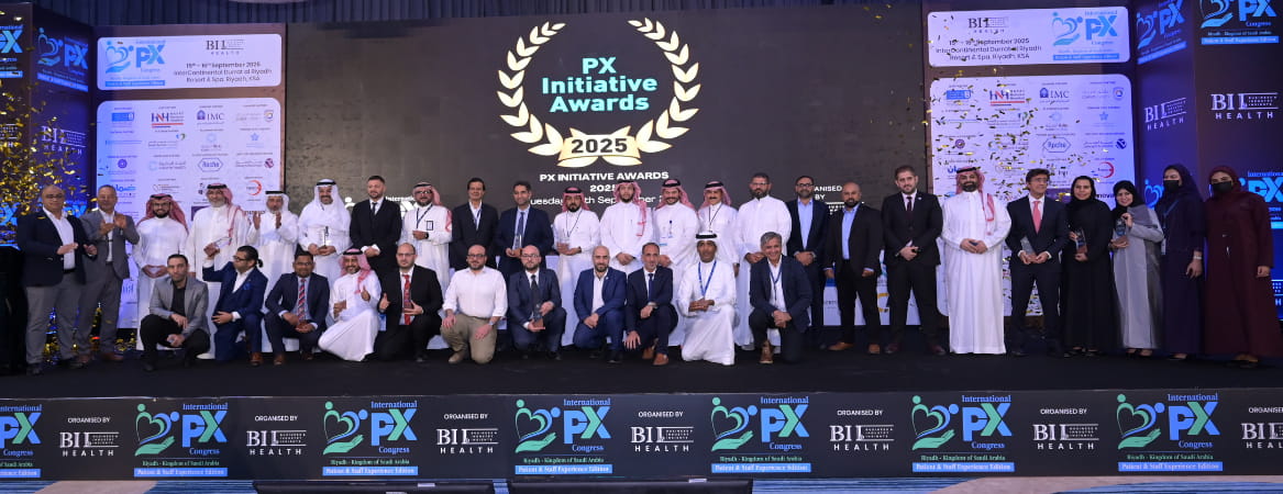
JHAH Wins International PX Congress Award for Patient Advocacy Excellence
September 28, 2025
JHAH’s award-winning patient advocacy project set a new global benchmark — cutting call light usage by 37% and boosting nurse response to 95%, delivering faster, safer care for every patient
JHAH Designated a Rezūm Center of Excellence for Non-Surgical Prostate Therapy
October 07, 2025
Breakthrough men’s health at JHAH. Rezūm therapy offers fast, non-surgical relief for enlarged prostate symptoms with proven results and faster recovery
Breast Cancer Survivor: ‘Screening Saved My Life’
October 12, 2025
Basmah’s story proves early detection saves lives. Book your screening today
Why Gulf countries need more 'patient champions'
October 20, 2025
Discover why empowering patients to share their health journeys is key to building a prevention-first healthcare culture across the Gulf.
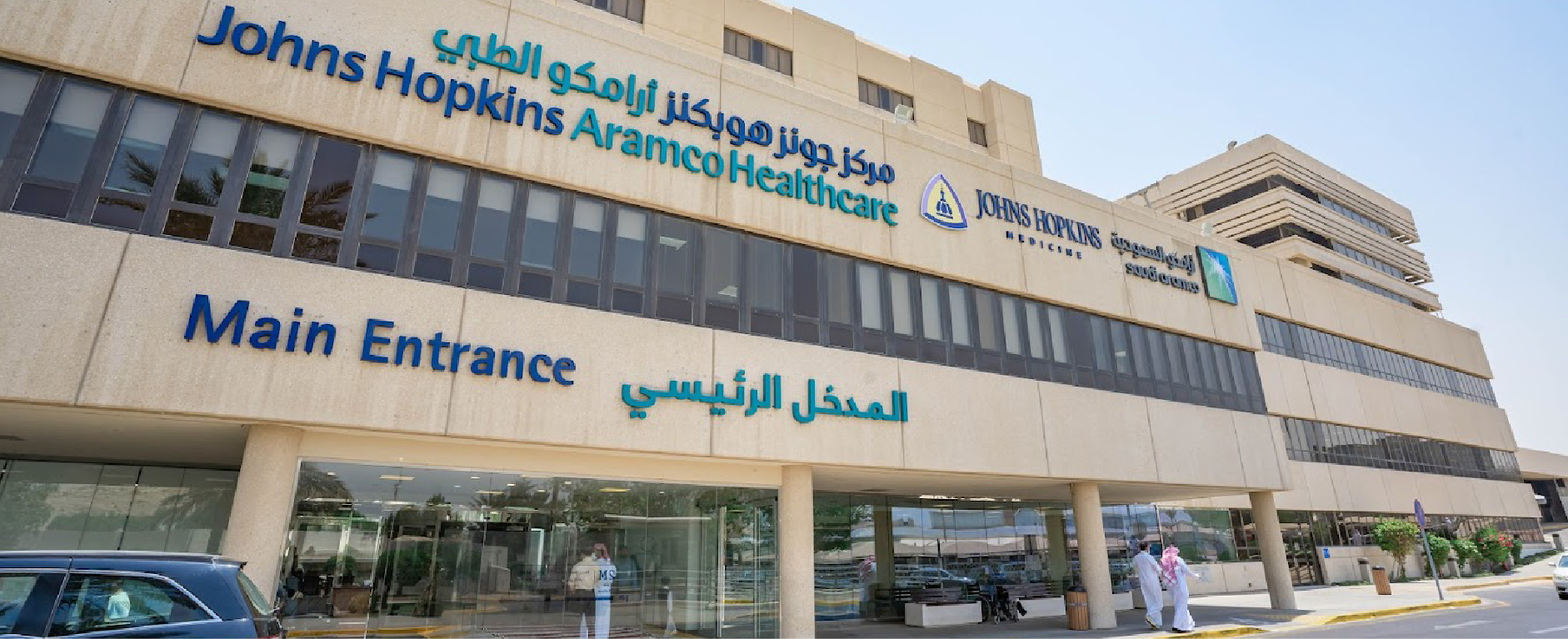
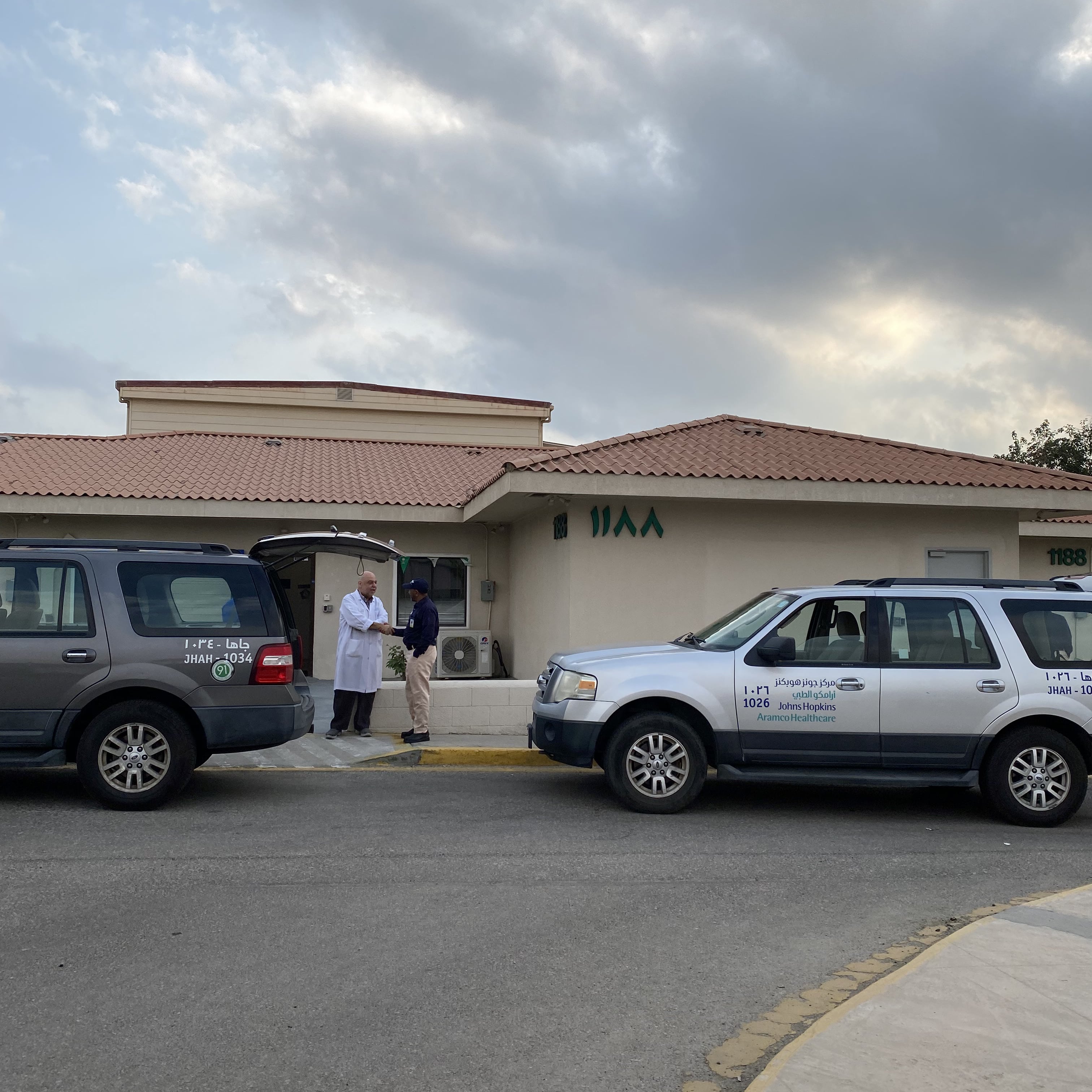
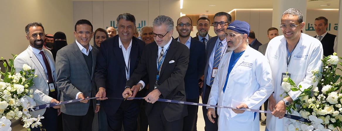
Johns Hopkins Aramco Healthcare opens doors to the public, marking a landmark moment in Saudi Arabia’s healthcare transformation
October 13, 2025
JHAH expands care to the public for the first time, marking a new chapter in Saudi Arabia’s Vision 2030 journey toward accessible, world-class healthcare.
Johns Hopkins Aramco Healthcare appoints Dr. Russell Hales to lead its Centers of Excellence, advancing specialized care in the Kingdom
October 15, 2025
Dr. Russell Hales, a Johns Hopkins Medicine expert, is appointed to lead JHAH’s Centers of Excellence, advancing integrated, world-class specialty care across Saudi Arabia.
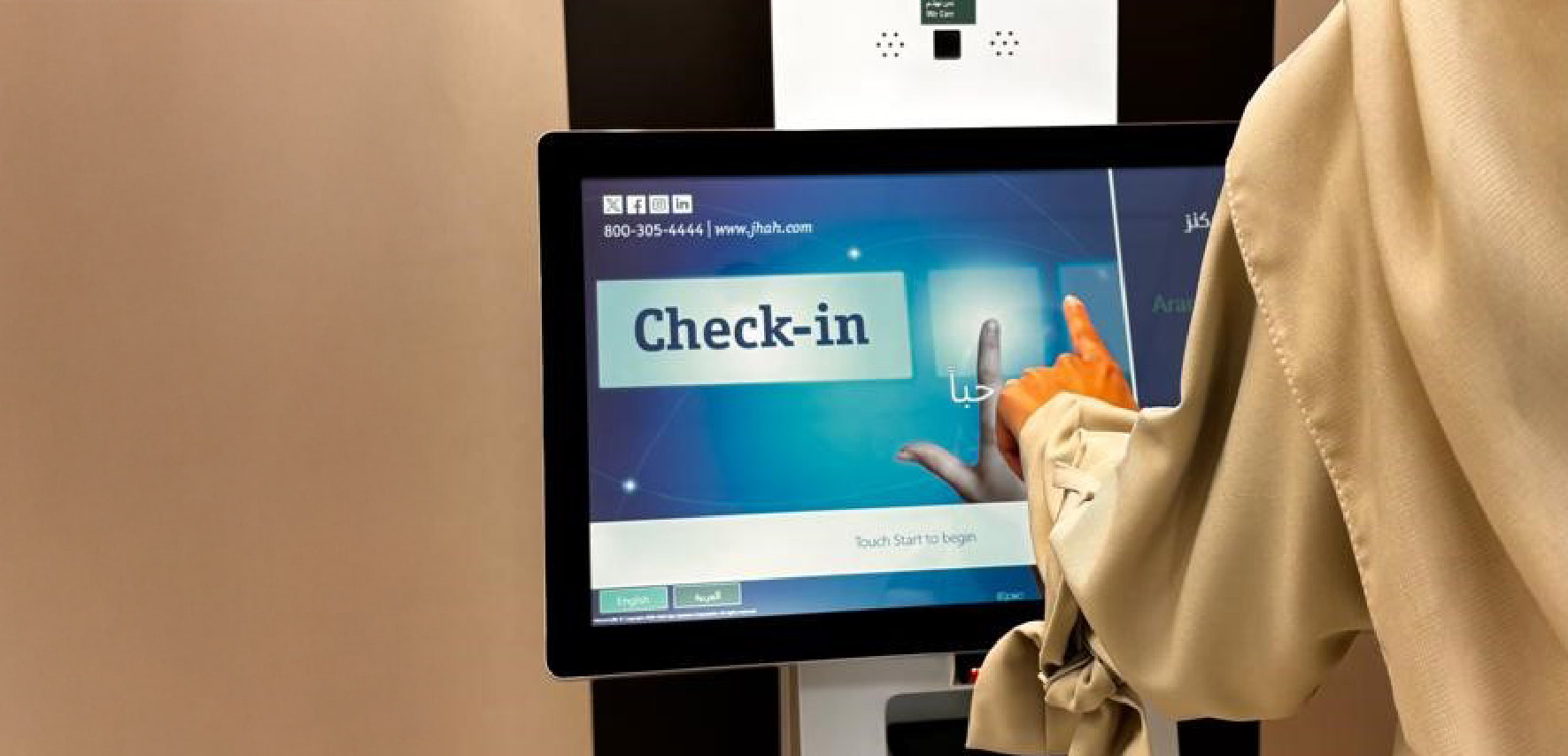
New Self-Service Check-In Kiosks
October 22, 2025
Check in quickly and easily using the kiosks at any JHAH outpatient specialty clinics, Primary Care and Dental receptions in Dhahran, Ras Tanura and Al-Hasa.
Robotic Surgery Program Expands to Include Thoracic Procedures
October 30, 2025
JHAH surgeons perform a first-of-its-kind robot-assisted thoracic procedure in Saudi Arabia, offering patients faster recovery and greater precision in minimally invasive care
Two JHAH Scientists Ranked in Global Top 2% by Stanford University
October 30, 2025
JHAH scientists Professors Ali Rabaan and Jaffar Al-Tawfiq were named among the world’s top 2% by Stanford University, recognizing their impact on global infectious disease research.
First Robotic Pediatric Surgery in Eastern Province
November 02, 2025
A new milestone in pediatric care: JHAH surgeons use robotic technology to deliver precise, minimally invasive treatment and faster recovery for children
American College of Surgeons Names JHAH As a Top Hospital for Surgical Care
November 16, 2025
The American College of Surgeons recognized JHAH as one of the region’s top hospitals for surgical safety, quality, and patient outcomes

JHAH Surgeons Save Patient’s Life with First Aortic Implant Procedure of its Kind in Kingdom
November 26, 2025
A JHAH team performed Saudi Arabia’s first TBE aortic implant, saving a patient’s life and setting a new benchmark for vascular surgery
Robotic Surgery Gives New Lease of Life to Ulcerative Colitis Sufferer
November 26, 2025
A robotic colorectal surgery at JHAH helped Ahmed regain his quality of life and finally put years of pain behind him

A JHAH Patient Speaks Out About Cervical Cancer Screening
December 18, 2025
Cervical cancer survivor Mariam Al Dossary explains how HPV screening at JHAH caught cancer early and why regular tests are a lifesaving “no‑brainer.”
JHAH Al-Hasa: First Baby-Friendly Hospital in Al-Hasa
January 01, 2026
First private hospital in Al-Hasa to receive Baby-Friendly Hospital accreditation from Saudi Ministry of Health.

Oncology Doctor, Majid Al Othman: Why I Chose Oncology
January 11, 2026
Radiation oncologist shares his inspiration, why he chose oncology, and the most memorable moments of his career.
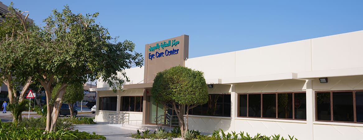
New Eye Care Center in Dhahran Now Open
January 19, 2026
Discover our newly opened Eye Care Center in Dhahran—modern, spacious, and equipped with advanced technologies for seamless, high‑quality eye care.
New Emergency Room Observation Area in Dhahran
January 27, 2026
Our expanded Emergency Room Observation Area is now open, offering a state‑of‑the‑art environment designed to enhance patient comfort, monitoring, and emergency care efficiency.
JHAH Launches Care Forward Plan with Eye Care Center
January 28, 2026
JHAH announces multi-year Care Forward Plan to optimize clinical infrastructure, beginning with expanded ophthalmology services.

JHAH Among First in Kingdom to Use Breakthrough Blood Cancer Drug
January 29, 2026
Cancer patients at Johns Hopkins Aramco Healthcare now have access to elranatamab, a new immunotherapy that gives hope to multiple myeloma patients who haven't responded to other treatments.

JHAH to Partner in Inaugural Applied Medical Research and Innovation Forum in Dammam
February 03, 2026
Johns Hopkins Aramco Healthcare supports the first Applied Medical Research Forum in Dammam, bringing together healthcare leaders to shape the future of medical innovation in the Kingdom.

Johns Hopkins Aramco Healthcare Earns Fourth Consecutive Mowa’amah Gold Standard Accreditation
February 23, 2026
JHAH has earned the Mowa'amah Gold Standard for the fourth consecutive term, reflecting a sustained commitment to accessible, dignified healthcare for persons with disabilities
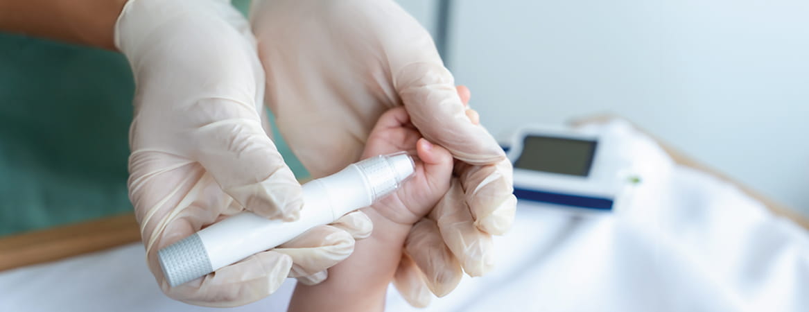
How JHAH's Emergency Team Detected Diabetes in an Infant and Changed One Family's Life
February 23, 2026
Reema's story shows how early detection of diabetes in an infant, made possible by JHAH's Dhahran emergency and pediatric teams, can be life-changing for the whole family
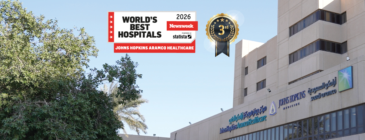
Johns Hopkins Aramco Healthcare Ranked 3rd Best Hospital in Saudi Arabia in Newsweek’s 2026 Report
February 26, 2026
Rising from 5th to 3rd in Saudi Arabia, JHAH earned its best-ever placement in the Newsweek World’s Best Hospitals 2026 ranking
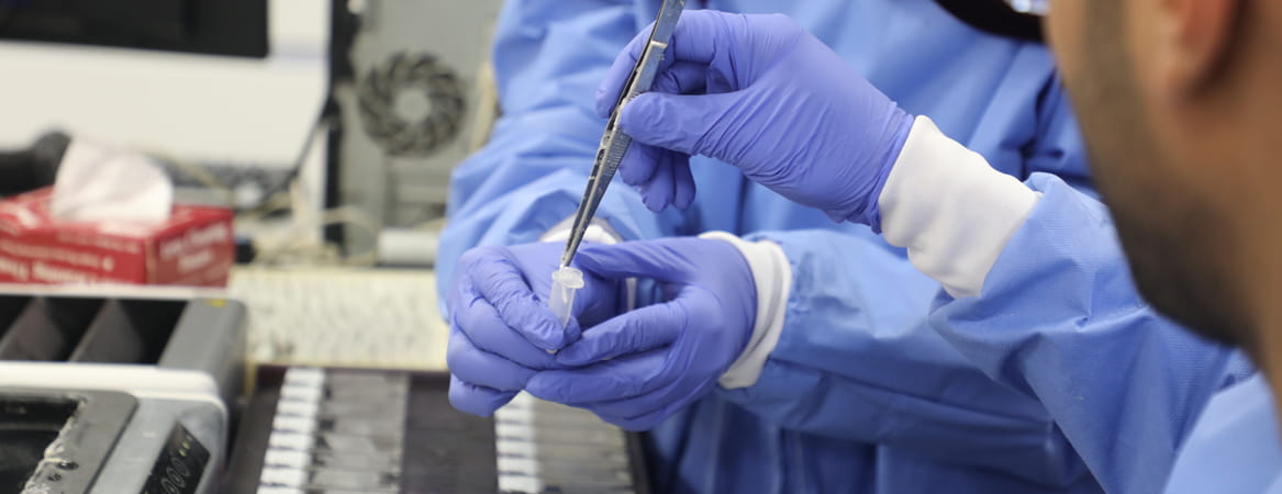
Introducing a New Era in Precision Diagnostics at JHAH: Our First In-house Molecular Oncology Test Result
March 11, 2026
Faster results, fewer referrals: JHAH's first in-house molecular oncology test strengthens our precision diagnostics and cancer care for patients in the region.